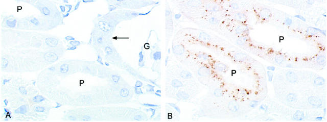
Figure 9.
Immunohistochemical analysis of lysozyme in megalin-deficient (A) and control kidney cortex (B). Cryosections from kidney cortex were incubated with rabbit anti-human lysozyme IgG followed by peroxidase-conjugated anti-rabbit IgG. Bound IgG was visualized by diaminobenzidine. In wild-type tissue (B), strong labeling for lysozyme is seen in apical endosomes and lysosomes of the proximal tubules (P). No staining is seen in megalin-deficient proximal tubules (A) including the very initial part of the tubule (arrow) that is connected to Bowman′s capsule of the glomerulus (G). Magnification, ×900.

