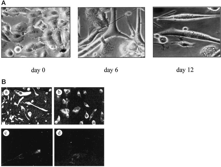
Figure 2.
Morphological and immunohistological analysis of MITC cells. Phase-contrast microscopy of MITC cells at passage 8 (A). After treatment with cytosine-arabinose the cells presented multiple nuclei, some fusing cells were seen at day 6, and small myotubes at day 12 were observed. B: In another cell preparation anti-desmin (a) and anti-troponin T (b) antibodies were clearly reactive with MITC, while anti-keratin antibodies were not (c). The control antibody was consistently negative (d).

