
Figure 4.
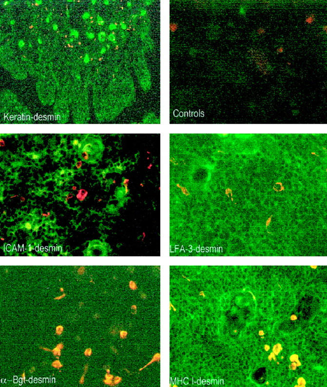
Analysis of myoid cells by double immunofluorescence on thymic sections. All microphotographs show superimposed staining of anti-desmin antibody (red staining) and one of the other antibodies (green staining). Double staining with anti-keratin antibody at a low magnification indicates that myoid cells (orange staining) are essentially located in the medulla and in the corticomedullary junction (magnification, ×40). Double staining with anti-ICAM-1 antibody indicates that myoid cells are mainly negative (magnification, ×200). Double staining with LFA-3 antibody shows a staining of the thymic network and indicates that myoid cells are positive (magnification, ×200). Double staining with α-Bgt indicates that myoid cells are the only cells that express α-Bgt binding sites (magnification, ×180). Double staining with anti-MHC class I antibody shows a strong staining of the thymic cells and indicates that myoid cells express MHC class I antigen (magnification, ×180).