
Figure 2.
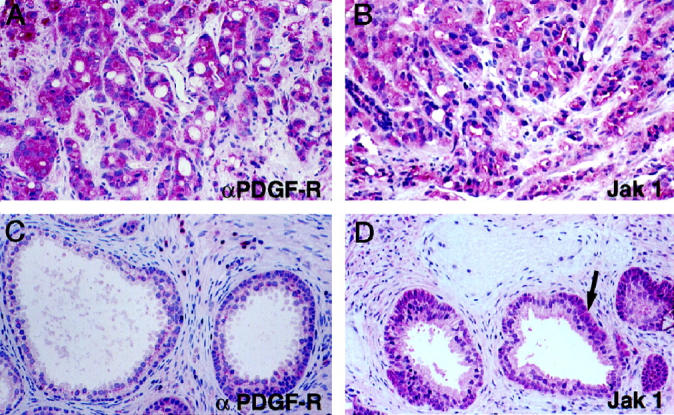
Immunohistochemical analyses of αPDGF-R and Jak 1 in primary PCa. A and B: Primary PCa stained with αPDGF-R and Jak 1 antibodies, respectively. C and D: Nonneoplastic prostate stained with αPDGF-R and Jak 1 antibodies, respectively. Arrow in D shows basal cell staining. Original magnification ×200.