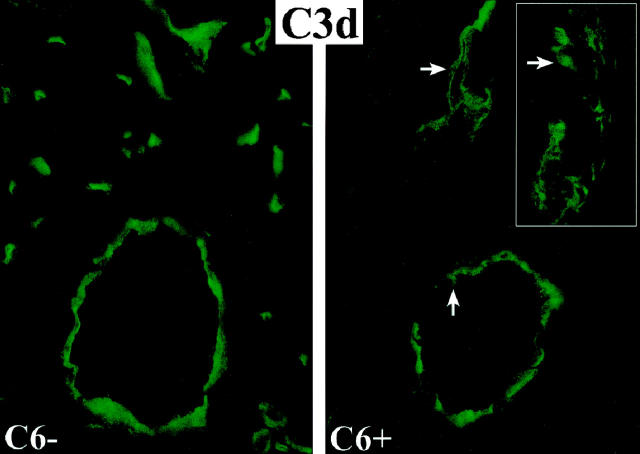
Figure 4.
Immunofluorescent stains for C3d on the endothelium of arteries, capillaries and veins in cardiac allografts to control (C6−) and C6 reconstituted (C6+) recipients. In the reconstituted C6+ recipients, subendothelial and perivascular depositions of C3d are evident in areas of vascular injury (arrows). The inset demonstrates at higher magnification the disruption of the endothelial cell layer (arrow) and deposition of C3d in the media of an artery.

