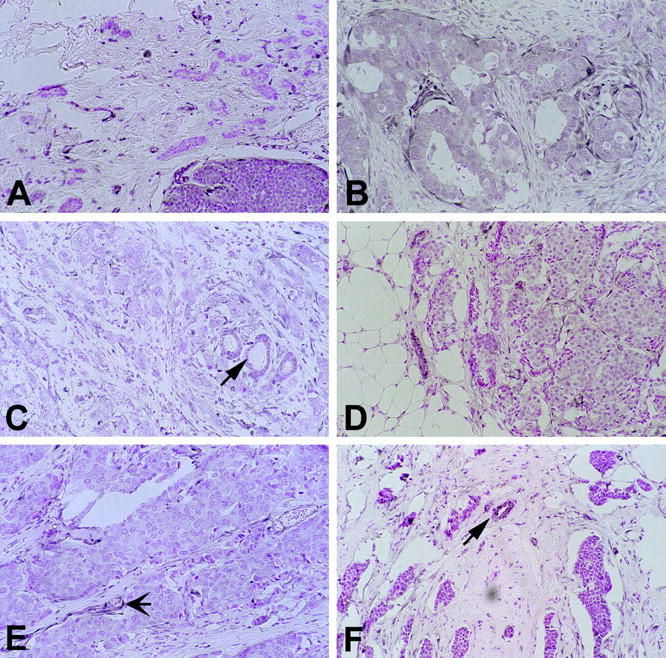
Figure 3.

Cases with weak staining (arrows in C and F, non-neoplastic duct; arrow in E, blood vessel). A: Ductal carcinoma (case 40) showing no staining (graded −) in the invasive component (top) adjacent to immunostain-positive intraductal component (bottom). B: Case 66. C: Case 59. D: Case 57. E: Case 45. Original magnification, ×30.
