
Figure 2.
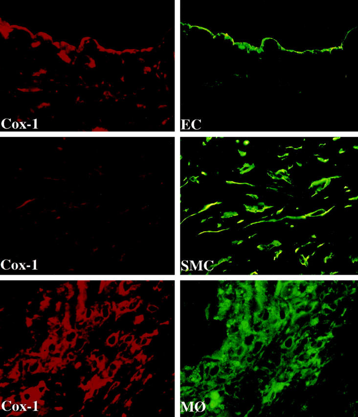
Colocalization of Cox-1 with endothelial cells (EC), smooth muscle cells (SMC), and macrophages (MΦ) in human atheroma. High power views (×400) of frozen sections of human carotid lesions showed specific staining for Cox-1 (red staining) on human vascular EC, SMC, and MΦ within the atheroma. Cell types were characterized by immunofluorescence-double labeling (green staining) as described in Materials and Methods. The lumen of the artery is at the top of each photomicrograph. Analysis of atheroma obtained from five different donors showed similar results.