
Figure 1.
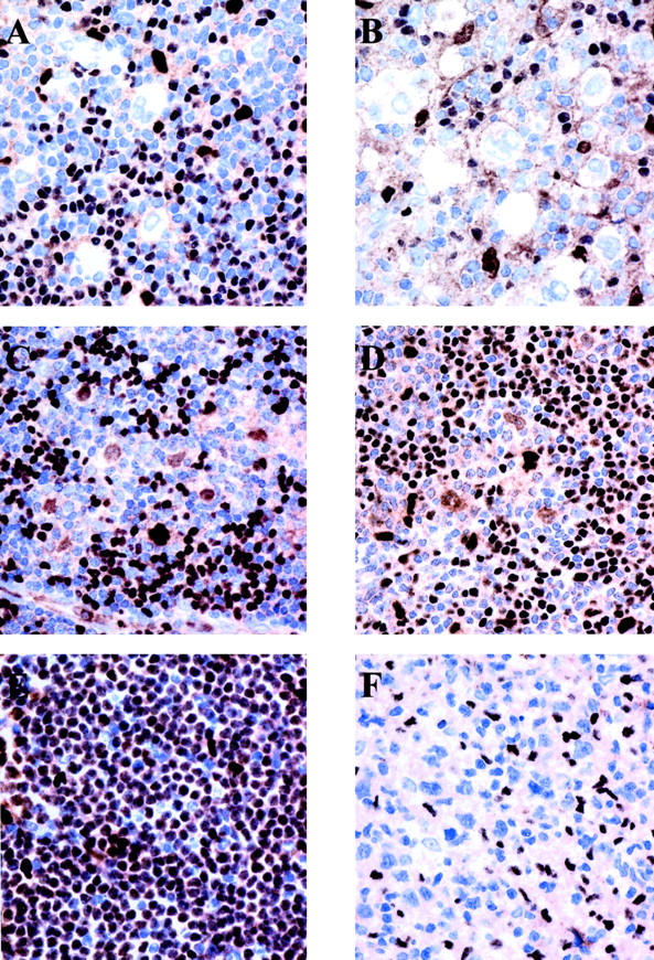
PU.1 is differentially expressed in HD and B-NHL. Immunohistochemical analysis on paraffin-embedded tissues using monoclonal anti-PU.1 antibody (immunostaining by the EnVision method using diaminobenzidine as a chromogen). A and B: Reed-Sternberg cells in nodular sclerosis cHD and mixed cellularity cHD, respectively, show no expression of PU.1. Small B lymphocytes and occasional histiocytes, but not T lymphocytes in the background express the protein in the nuclei. C and D: Neoplastic cells in two cases of LPHD expressing PU.1 in the nucleus. T cells rosetting the neoplastic cells do not express PU.1, whereas small B cells and histiocytes in the background do express the protein. E: PU.1 is strongly and diffusely expressed in small lymphocytic lymphoma/chronic lymphocytic leukemia. F: Absence of PU.1 expression by the neoplastic cells of DLBCL.