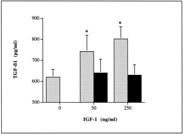
Figure 8.
Insulin stimulation of TGF-β1 was mimicked by the addition of recombinant IGF-1. HK-2 cells were stimulated with increasing doses of IGF-1 (0 to 250 ng/ml) under serum-free conditions in the presence (filled bars) or absence (shaded bars) of 50 ng/ml of the monoclonal antibody to IGF1R (B). Supernatant samples were collected for determination of TGF-β1 after 48 hours. Data represents mean ± SD (n = 8; *, P < 0.05 versus unstimulated control and IGF stimulated in the presence of antibody).

