Figure 3.
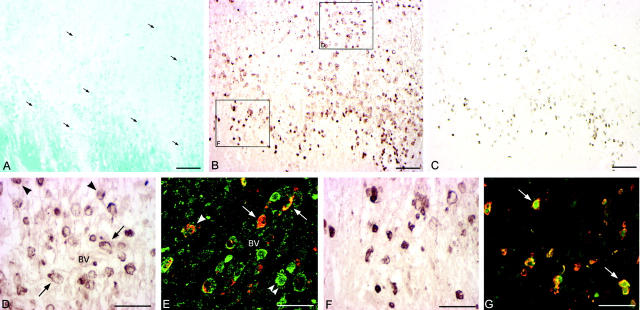
Characterization of CCR1 expression in EA region and expanding lesion edge of actively demyelinating MS lesion. Actively demyelinating and expanding lesion stained with luxol fast blue myelin stain (LFB, A) indicating the lesion edge and adjacent EA area. Macrophages contained myelin degradation products indicating ongoing demyelination (A, arrows). Immunohistochemistry for CCR1 (B, D, and F) located CCR1+ cells to the immediate lesion edge and within EA regions. CCR1+ cells within EA areas were either perivascular (D, arrows) or dispersed in the parenchyma (D, arrowheads). Dual-label immunohistochemistry for CCR1 and CCR5 (E, CCR1 immunoreactivity shown in red, green indicates CCR5 immunoreactivity) and confocal microscopy characterized perivascular (E, arrows) as well as parenchymal (E, arrowhead) CCR1+/CCR5+ cells. Interestingly, all CCR1+ cells co-expressed CCR5, whereas CCR5+ cells not necessarily were also CCR1+ (E, double arrowheads). CCR1+ cells at the lesion edge (F) had the morphology of small, round cells. In a serial section analysis these cells co-localized with MRP14 immunoreactivity (C). Dual-label immunohistochemistry confirmed co-expression of CCR1 with MRP14 as shown in G (arrows, CCR1 immunoreactivity shown in red, green indicates MRP14 immunoreactivity). Scale bars: 100 μm (A–C), 50 μm (D–G). BV, blood vessel.

