
Figure 2.
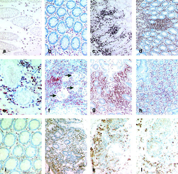
Localization of Lptn and GPR-5 expression in CD. a–h: Immunohistochemistry and mRNA in situ hybridization for Lptn. In control tissue samples of the large bowel Lptn mRNA (a) and protein (b) expression was found to be restricted to a few lymphocytes within the lamina propria. In tissue specimens affected by CD increased numbers of cells expressing Lptn mRNA (c) and protein (d) were found predominantly within lymphocytic aggregates of the lamina propria and submucosa. Within the subserosa Lptn-positive cells were predominantly found in a perivascular pattern (e, immunohistochemistry). Giant cells (arrows) or epithelioid cells of granulomas did not show any Lptn protein expression in contrast to surrounding T cells (f). Biopsy specimens obtained from a patient with active CD (severity activity index) demonstrated an increased number of Lptn-positive cells (g, immunohistochemistry), which were found to be significantly reduced on therapy with systemic steroids (severity activity index, 35) (h, immunohistochemistry). i–l: In situ hybridization for GPR-5 mRNA. In noninflammatory-affected bowel the expression of GPR-5 mRNA was restricted to cells within the lamina propria (i). In CD an increased number of GPR-5 mRNA-positive cells were found within the lamina propria (j), whereas cells with the strongest intracytoplasmic expression for GPR-5 mRNA were found adjacent to submucosal vessels (k) and granulomas (l). Original magnifications: ×100 (c, d, g, h, j); ×200 (a, b, f, i, k, l); ×400 (e).