Figure 4. .
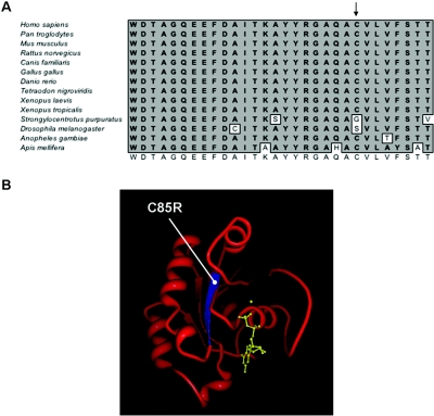
Sequence conservation and structural context of C85R substitution. A, Amino acid sequence comparison of the Switch 2 region of human RAB23 (top) with 13 other species. The consensus sequence is shown at the bottom, and the position of the mutated C85 residue is indicated with an arrow. B, Structure of human RAB2314 (Protein Data Bank [number 1Z22]), showing the C85 residue located in a β-strand (blue) and completely buried in the core of the protein. The bound Mg-GDP is shown in yellow. The structure was modeled using the Protein Workshop tool (Protein Data Bank).

