
Figure 3.
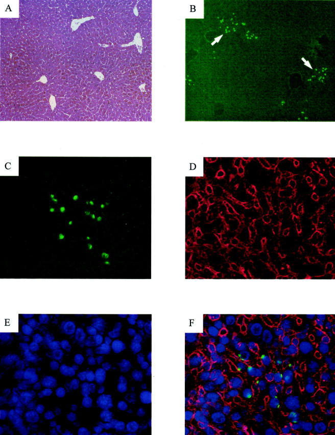
Histology and fluorescent in situ hybridization of a Bcl-xL-repopulated liver. A: Liver section examined by routine hematoxylin and eosin staining. Original magnification, ×100. B: Y chromosome staining in a liver section of the same animal. Arrows indicate repopulating-hepatocyte clusters. Original magnification, ×100. C–F: Identical fields showing a detailed view of a hepatocyte cluster. Original magnification, ×400. C: Staining for the Y chromosome FISH (green). D: Immunostaining with an antibody to cytokeratins 8, 18, and 19 (red). E: DAPI nuclei staining (blue). F: Overlays of the Y chromosome, cytokeratins and DAPI fluorescence.