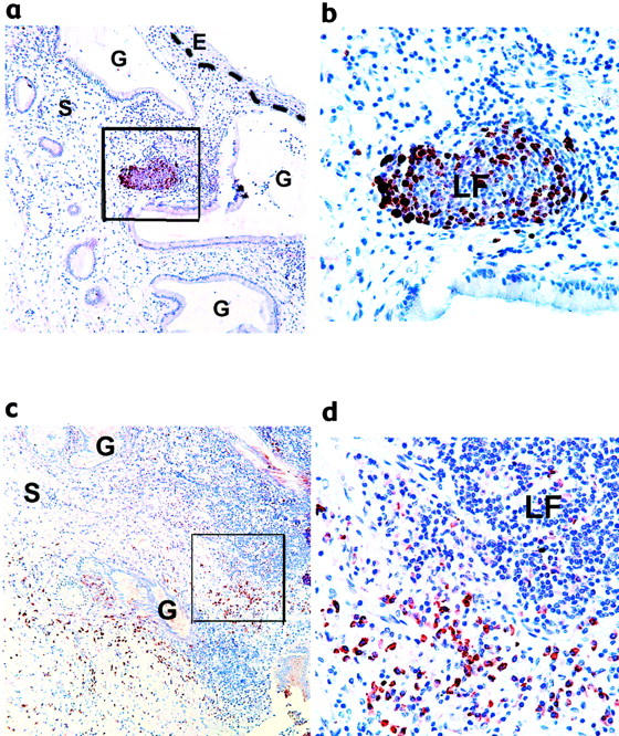
Figure 3.

Functional characterization of cervical GCs. Immunohistochemistry on paraffin-embedded sections of high-grade SIL with GC and LF-like structures was performed with antibody against Ki67 (a and b) and BCA-1 (c and d). The stromal-epithelial junction is marked with a dashed line. Photos were taken with the 10X (a and c) or 40X objective (b and d). b and d are the areas outlined in a and c, respectively. Immunoreactivity toward mouse IgG1 (isotype control) was negligible (data not shown). E, epithelium; S, stroma; G, endocervical gland; LF, lymphoid follicle.
