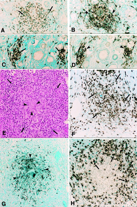
Figure 3.

Ovarian inflammation in pZP3(mac)- and pZP3(hu)-immunized macaques. A–D: Adjacent sections of a typical primate AOD lesion consisted of clusters of CD3+ T cells (A, arrows) and MHC II + cells (B, arrowheads). C and D: Adjacent sections of another typical primate AOD lesion stained for CD3 (C) and MHC II (D). Adjacent sections of a large granuloma in the ovary of Eenie that was immunized with pZP3(hu) in adjuvant. E: Typical granulomatous inflammation with eosinophilic center (arrowheads) surrounded by cuff of lymphocytes (arrows) (H&E). The granuloma contains many macrophage (Mac 387+) (F), a central core of MHC II-positive cells (G), and a rim of CD3+ T cells (H). Original magnifications, ×400.
