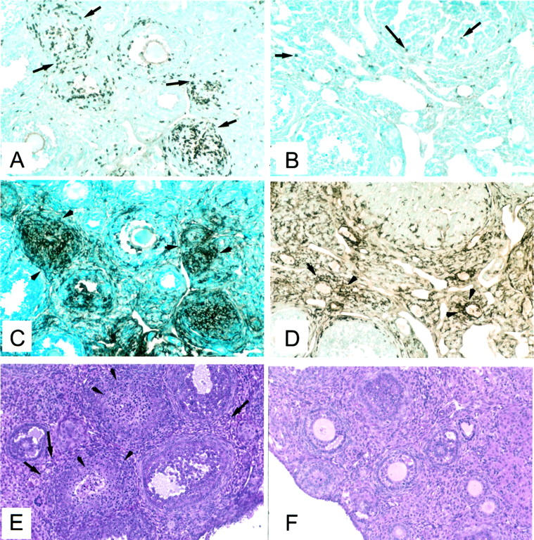
Figure 5.

Immunohistopathology of murine AOD. A, C, and E: Ovary of a mouse immunized with pZP3(mou) in CFA. Adjacent sections showing clusters of cells T cells (CD5+) (A, arrows) that co-localize with intensely MHC II+ cells (C, arrowheads). The opposite ovary stained with H&E (E) shows lymphocytic infiltration in the interstitial regions (arrows) and the ovarian follicles (arrowheads). B, D, and F: Ovary of an adjuvant-treated mouse showing a few T (CD5+) cells (B) and an adjacent section (D) with many MHC II+ macrophages in the interstitium and the corpus luteum. The opposite ovary (F) stained with H&E appears completely normal.
