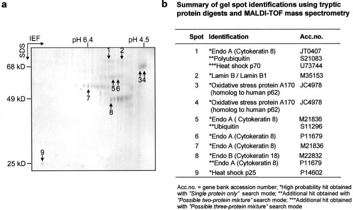
Figure 1.
Analysis of MB components by MALDI-TOF mass spectrometry. a: Coomassie blue-stained 2D gel of MB proteins isolated from DDC-intoxicated mouse liver. Numbers indicate protein spots used for MALDI-TOF analysis. b: Summary of gel spot identifications using tryptic protein digests and MALDI-TOF mass spectrometry.

