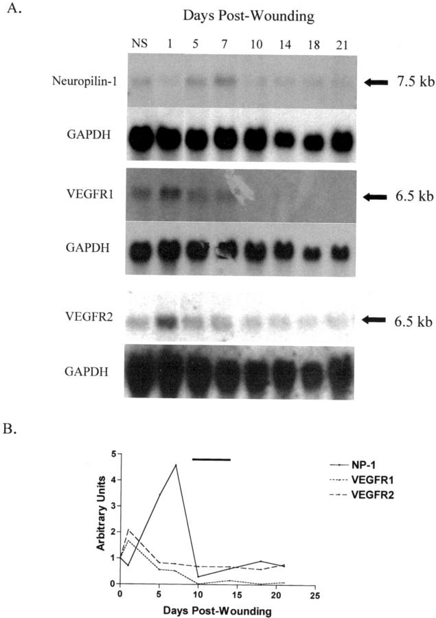
Figure 1.
VEGF receptor mRNA expression in wounds. A: Neuropilin-1, VEGFR1, and VEGFR2 mRNA levels were analyzed in normal skin and wound samples isolated at selected times from day 1 to 21 after wounding. Neuropilin-1 mRNA (7.5-kb band) was observed to reach maximal levels at day 7. In contrast, VEGFR1 and VEGFR2 mRNA (each represented by a 6.5-kb band) reached peak expression at day 1. The Northern blot for neuropilin-1 is representative of seven experiments. The Northern blots for VEGFR1 and VEGFR2 were performed in duplicate. GAPDH was used as a control for RNA loading. NS, normal uninjured skin. B: The mRNA expression pattern for each receptor was assessed by densitometry of the Northern blots depicted in (A). For each receptor, mRNA levels were normalized to GAPDH and the values are relative to those of normal skin. The bar above the graph represents the peak of angiogenesis in this model.

