
Figure 11.
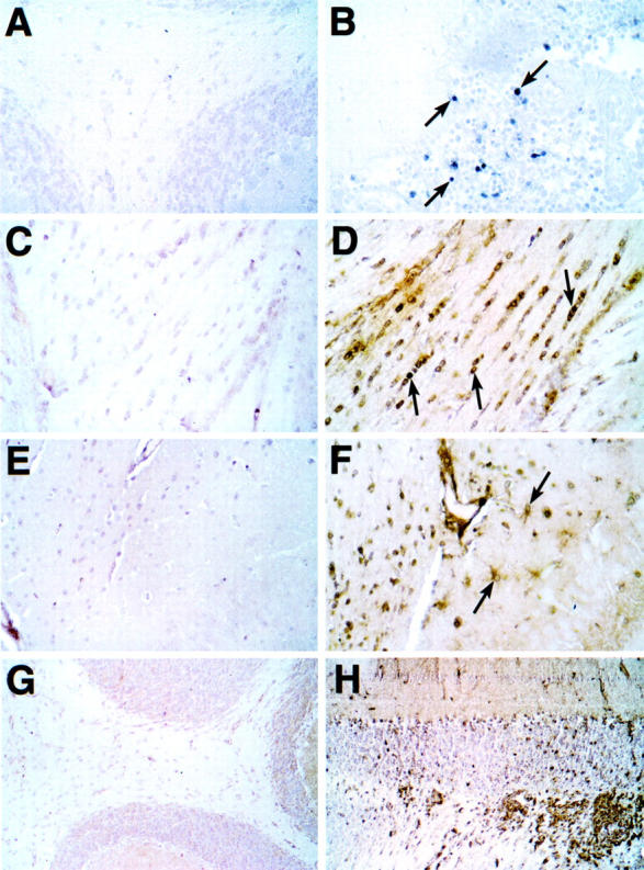
Distribution of STAT4 and STAT1 proteins in the brain during EAE. Sections from Bouin’s-fixed brain were immunostained with polyclonal antibody against either murine STAT4 (A and B) or STAT1 (C–H) protein as described in Materials and Methods. Although little staining was detectable for STAT4 in brain from control mice (A), numerous mononuclear cells were seen with strong cytoplasmic and nuclear staining in infiltrates in cerebellum (B, arrows) and spinal cord from mice with EAE. Very low staining for STAT1 was observed in brain from control mice (C, E, and G). In brains from mice with EAE, STAT1 staining was increased dramatically in most regions of the brain including subcortical periventricular white matter tracts (D), hippocampus (F), and cerebellum (H). In white matter nuclei with the morphological and spatial characteristics corresponding to oligodendrocytes showed strong nuclear staining for STAT1 (D, arrows). Cytoplasmic staining of cells with the morphological characteristics of astrocytes (F, arrows) is shown in the hippocampal region. Original magnifications: ×400 (A–F); ×100 (G and H).