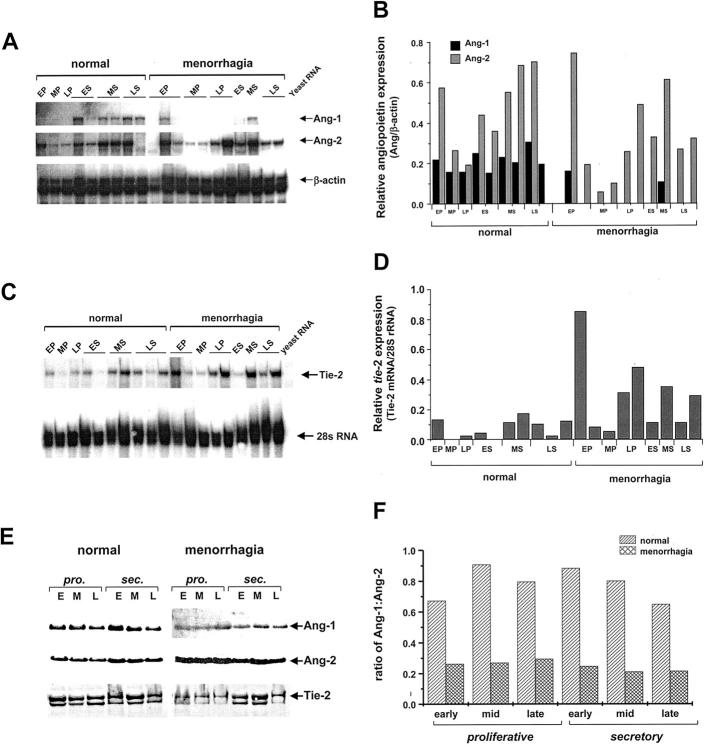
Figure 1.
Quantitative analysis of Ang-1, Ang-2, and Tie-2 expression in normal and menorrhagic endometrium. A: A representative RNase protection autoradiograph showing the relative expression of Ang-1 and Ang-2 mRNA expression in normal 9 and menorrhagic 11 endometrium samples. Endometrial samples were collected at the early proliferative (EP), mid-proliferative (MP), late proliferative (LP), early secretory (ES), mid-secretory (MS) and late secretory (LS) phases of the menstrual cycle. Total RNA was extracted from endometrial tissue and analyzed by RNase protection for the presence of Ang-1, Ang-2, and β-actin (control) mRNA. B: Densitometric analysis was performed on the autoradiographs and the densities of the Ang-1 and Ang-2 protected fragments normalized to the internal β-actin control fragments and represented graphically as relative density units. C:Tie-2 expression in endometrium was determined by RNase protection and the protected Tie-2 fragments normalized to 28s rRNA. D: The relative levels of Tie-2 mRNA in normal 9 and menorrhagic 11 endometrium samples is represented graphically as relative densitometric units. E: Ang-1, Ang-2, and Tie-2 protein expression was examined in endometrial samples by Western blot analysis. Protein extracts (50 μg) pooled from endometrial tissue from 5 individuals taken at early (E), mid- (M), and late (L) proliferative (pro.) and secretary (sec.) phases of the menstrual cycle were subjected to Western blotting and enhanced chemiluminescent detection with antibodies against Ang-1, Ang-2, and Tie-2. F: Densitometric analysis of Ang-1 and Ang-2 expression was performed on the luminographs and the results expressed as the ratio of Ang-1:Ang-2 for normal and menorrhagic endometrium throughout the cycle.

