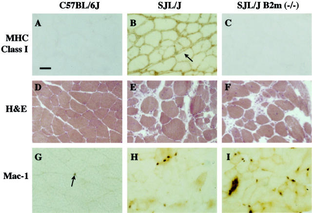
Figure 1.
A–C: MHC class I expression is up-regulated in muscles of SJL/J mice compared to muscles of SJL/J B2m (−/−) and C57BL/6J mice. A: Immunohistology of C57BL/6J gastrocnemius muscle showed little staining, a result consistent with the very low expression of MHC class I proteins on normal muscle fibers. B: In contrast, SJL/J muscle showed dark staining apparently on the surface of muscle fibers (eg, arrow), as well as staining on cells in between, and sometimes within, muscle fibers. C: In contrast, SJL/J B2m (−/−) muscle showed very little staining, a result expected for tissues lacking β-2-microglobulin. Scale bar, 40 μm. D–F: Muscles from SJL/J and SJL/J B2m (−/−) mice showed similar extents of myopathy. D: H&E staining showed normal fibers in wild-type C57BL/6J gastrocnemius muscle. E and F: In contrast, abnormal fibers were abundant and found at similar percentages in SJL/J (E) and SJL/J B2 m (−/−) (F) muscles as described in Table 1 ▶ . G and H: Mac-1-positive cells were most abundant in myopathic muscles of SJL/J B2m (−/−) mice, moderately abundant in SJL/muscles, and rare in the normal muscles of C57BL/6J mice. G: Immunohistochemistry showed that Mac-1-postive cells (arrow) were rare in the gastrocnemius muscle of C57BL/6J wild-type mice. H and I: In contrast, Mac-1-positive cells were abundant in SJL/J (H) and SJL/J B2m (−/−) (I) muscles, being most abundant in the B2m (−/−) muscles as described in Table 2 ▶ .

