
Figure 1.
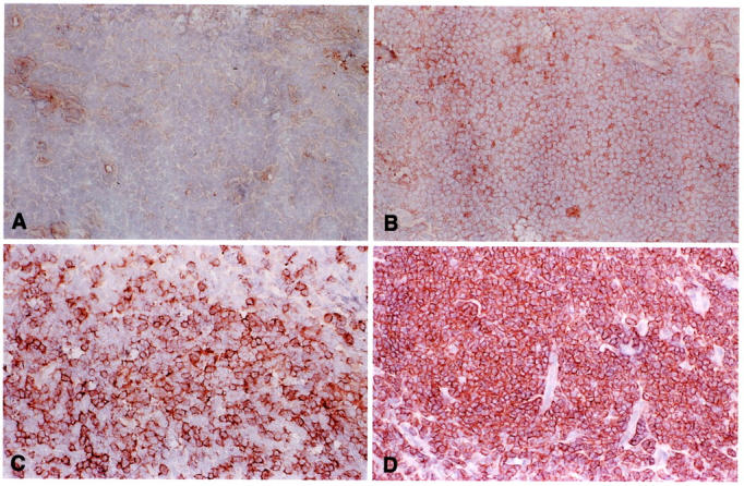
FL 8-‘83 harbors both IgM- and IgG-expressing tumor cells. Frozen section of FL 8-‘83 stained with monoclonal antibodies specific for human IgA (A), IgG (B), IgM (C), and BCL-2 (D) (magnification, ×100). Whereas all cells are IgA-negative, a faint but significant expression of IgG is found on the majority of cells in all areas. In between these cells, scattered tumor cells with relatively strong IgM expression are found.