Figure 2.

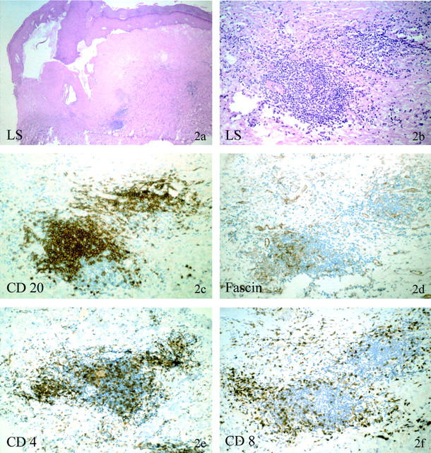
Immunohistochemical phenotype of deep dermal/submucosal lymphocytic aggregates in atrophic LS with monoclonal γ-TCR rearrangement. Patient no. 21 (30665/99). The H&E stain shows a late stage of LS with massive hyperkeratosis and focal acanthotic epidermis. The basement membrane is massively thickened, the papillary dermis highly sclerotic and avascular with prominent clefting along the dermoepidermal junction. No lichenoid dermal or interface infiltrate is present; however, beneath the sclerosis several lymphocytic cell aggregates are present (a and b). These aggregates are composed of predominately CD20-positive B cells (c), fascin-positive dendritic cells (d), and CD4-positive T cells (e). CD4-positive T cells dominate over CD8-positive T cells (f), which are not found within the B-cell fraction but at the periphery of these lymphocytic aggregates.
