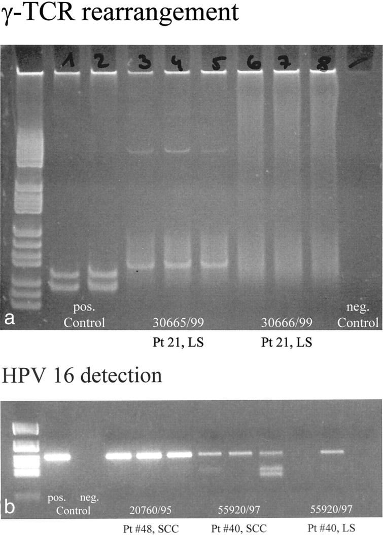
Figure 3.

PCR analysis of the lymphoid infiltrate of LS for γ-TCR rearrangement. a: Gel electrophoretic analysis with positive controls in lanes 1 and 2, and negative controls in lane 9. Lanes 3 to 5 represent PCR products of three different areas of a biopsy of LS (patient no. 21, 30665/99) of an 89-year-old female that show a single band in all three lanes corresponding to monoclonal rearrangement of the γ-TCR. The same analysis (lanes 6 to 8) of a different lesion biopsied in the same patient (patient no. 21, 30666/99) at the same time shows no bands, indicating the absence of monoclonal γ-TCR rearrangement. PCR analysis of LS for the presence of HPV16 DNA. b: The detection of HPV16 DNA on an ethidium bromide gel with positive and negative controls in lanes 1 and 2. Lanes 3 to 5 are a typical example of detection of strong bands of HPV16 DNA in all three samples obtained from a single paraffin block (patient no. 48, SCC without LS). In another patient with SCC arising in LS (patient no. 40), weaker bands of HPV16 DNA are visualized in all three samples of the analyzed block containing SCC in lanes 6 to 8. Lanes 9 to 11 illustrate the typical finding of HPV16 DNA in LS as a weak band in just one of three samples of an analyzed biopsy of LS (patient no. 40).
