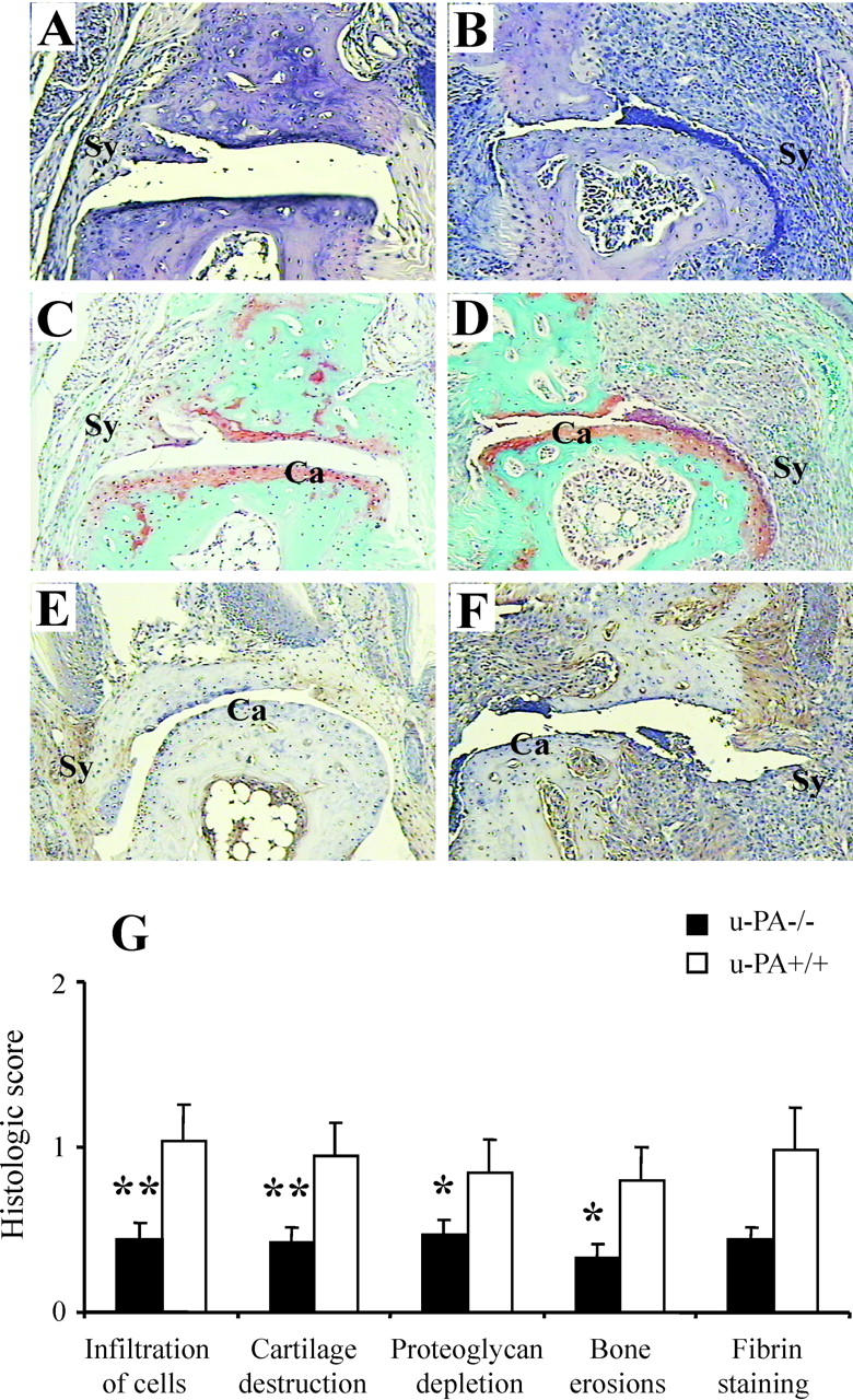
Figure 3.

Histological assessment of CIA in u-PA−/− mice. Distal interphalangeal joints from u-PA−/− mice (A, C, and E) and u-PA+/+ mice (B, D, and F). These are representative of average clinical scores in each case. A and B: H&E staining. C and D: Safranin O, fast green staining with a hematoxylin counterstain. E and F: Immunohistochemical detection of fibrin. Note that the arthritis is milder in the joints from the u-PA−/− mice and there is only very weak fibrin staining. Ca, cartilage; Sy, synovium. Original magnifications, ×125. G: Histological scores (mean ± SEM) for each histological feature (n = 26 for u-PA−/− and 18 for u-PA+/+ limbs) and for fibrin staining (n = 10 for u-PA−/− and 10 for u-PA+/+ limbs). Note that scoring for fibrin staining is 0 to 6 and for all other histological features is 0 to 3. *, P < 0.05; **, P < 0.01 u-PA−/− versus u-PA+/+ mice.
