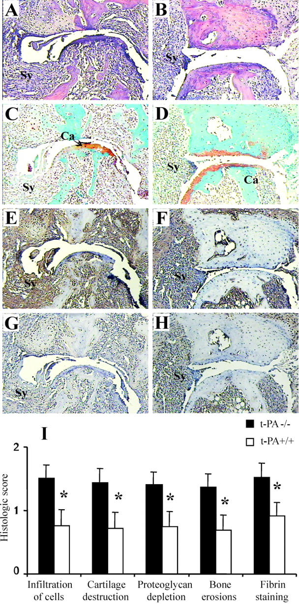
Figure 4.

Histological assessment of CIA in t-PA−/− mice. Severely affected metatarsal phalangeal joints from t-PA−/− mice (A, C, E, and G) and t-PA+/+ mice (B, D, F, and H). These represent joints with the most severe arthritis in each case. A and B: H&E staining. C and D: Safranin O, fast green staining with a hematoxylin counterstain. E and F: Immunohistochemical detection of fibrin. G and H: Control staining for fibrin. Note that the arthritis is more severe in the joint from the t-PA−/− mouse and there is more fibrin staining. Ca, cartilage; Sy, synovium. Original magnifications, ×125. I: Histological scores (mean ± SEM) for each histological feature (n = 14 for t-PA−/− and 15 for t-PA+/+ limbs) and for fibrin staining (n = 11 for t-PA−/− and 9 for t-PA+/+ limbs). Note that scoring for fibrin staining is 0 to 6 and for all other histological features is 0 to 3. *, P < 0.05 t-PA−/− versus t-PA+/+ mice.
