
Figure 5.
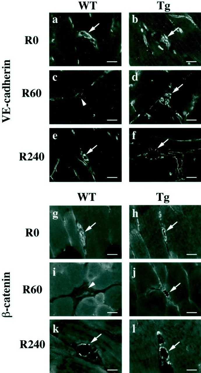
Endothelial adherens junction assembly in reperfused vessels. Immunofluorescence of adherens junction components VE-cadherin (a–f) and β-catenin (g–l) are shown. Immunoreactivities of VE-cadherin and β-catenin were detected using a goat polyclonal anti-VE-cadherin antibody and a mouse monoclonal anti-β-catenin antibody. After reperfusion for 60 minutes, both VE-cadherin and β-catenin disappeared from cell-cell contacts of endothelial cells in reperfused vessels in WT mice (arrowhead, c, i). On the other hand, this disappearance was not seen in eNOS-Tg mice at the same time point (arrow, d, j). After 240 minutes of reperfusion, adherens junction components were localized at cell-cell contacts in endothelial cells in both WT (e, k) and eNOS-Tg mice (f, l). The figures are representatives of WT (n = 6) and eNOS-Tg (n = 6) mice. Similar results were obtained from all of the examined mice of both genotypes. Scale bar, 20 μm.