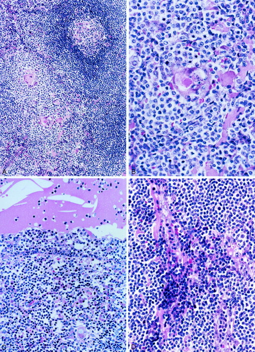
Figure 3.

Histological appearance of thymic MALT lymphoma. A: A lymphoid follicle with intact mantle zone is surrounded by a diffuse small lymphoid infiltrate. B: Lymphoepithelial lesion formed by a Hassall’s corpuscle infiltrated by CCL cells. C: The epithelium lining the cyst is invaded by CCL cells forming the lymphoepithelial lesion. The cyst contains eosinophilic material and cholesterol crystals. D: Mature plasma cells are found around the vessels that show mild sclerosis. H&E; original magnifications: ×40 (A), ×100 (B), ×66 (C), ×80 (D).
