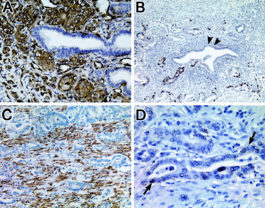
Figure 2.

Validation of gene expression by immunohistochemical and in situ hybridization in primary pancreatic cancers. A: Fascin. Strong cytoplasmic immunolabeling is noted within the infiltrating neoplastic epithelium, in contrast to the normal pancreatic duct epithelium that is negative. B: Topoisomerase IIα. Strong nuclear immunolabeling is noted within the neoplastic epithelium, in contrast to the normal pancreatic duct epithelium (black arrows) and desmoplastic stroma that are negative. C: Heat shock protein 47. Strong immunolabeling is noted of the desmoplastic stroma of the tumor, in contrast to the neoplastic epithelium that is negative. D: Pleckstrin. mRNA expression is detected within the neoplastic epithelium by in situ hybridization (black arrows), in contrast to the surrounding desmoplastic stroma that is negative. The nonneoplastic epithelium also did not label.
