
Figure 9.
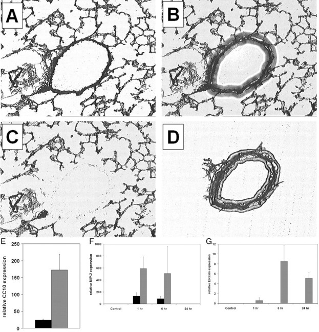
LCM and TaqMan PCR analysis of bronchiolar epithelium and alveolar epithelium following a single nebulized allergen challenge. Mice received an intraperitoneal injection of OVA at days 1 and 14 and were nebulized with aerosolized OVA at day 21. Control mice were sensitized but not exposed to aerosolized OVA. Mice were euthanized at various times following a single allergen challenge, and frozen lung sections were prepared. Sections were visualized by light microscopy (A), a transfer cap was placed atop the section and a laser was pulsed, causing the film to melt and adhere to the section (B). The cap was lifted away, revealing the remaining tissue (C) and the enriched fraction on the cap (D). Bronchiolar epithelial (gray bars) and parenchymal tissues (black bars) were laser captured from mouse lung sections and the RNA expression of the bronchiolar epithelial specific gene product, CC10, was quantitated relative to the expression of the housekeeping gene 18S ribosomal RNA using TaqMan PCR (E). Quantitation of MIP-2 mRNA (F) and eotaxin mRNA (G) expression in bronchiolar epithelium and parenchymal tissue of sensitized mice following a single aerosolized allergen challenge. mRNA levels are normalized to 18s rRNA. Data are from four mice per group and are expressed as means ± SEM.