
Figure 1.
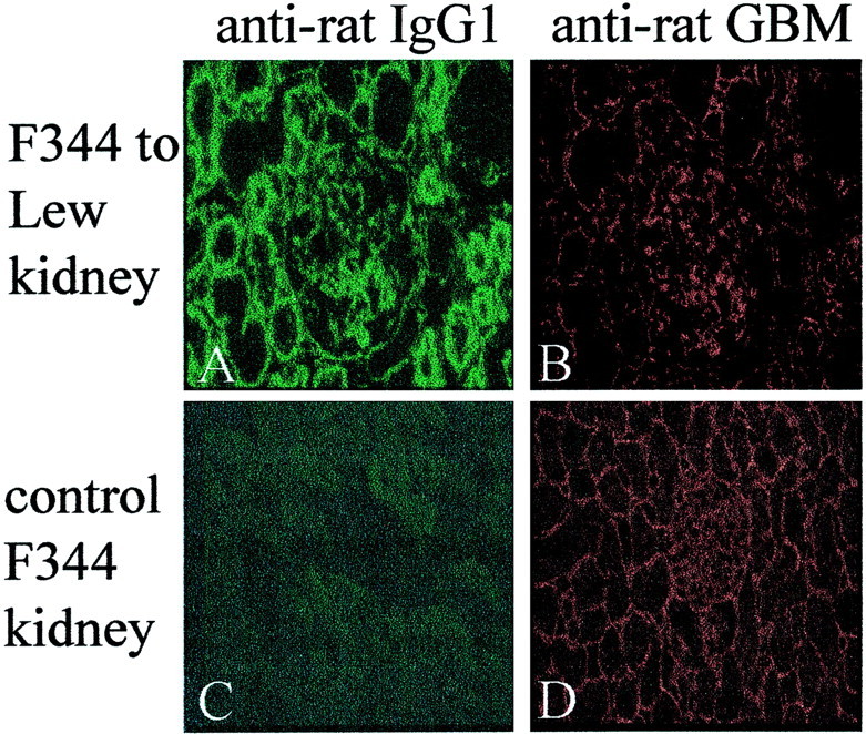
Immunofluorescent staining for in vivo immunoglobulin deposition of antibodies after Tx. A: F344 kidney graft removed on day 60 after Tx from a LEW recipient and stained for rat IgG1. B: Same kidney section (A) incubated with a rabbit anti-rat GBM antiserum and stained for rabbit IgG. C: Normal F344 kidney section stained for rat IgG1. The staining is similar to LEW kidney grafts removed from F344 recipients. D: Normal F344 kidney section incubated with rabbit anti-rat GBM antiserum and stained for rabbit IgG. Original magnifications, ×250.