
Figure 9.
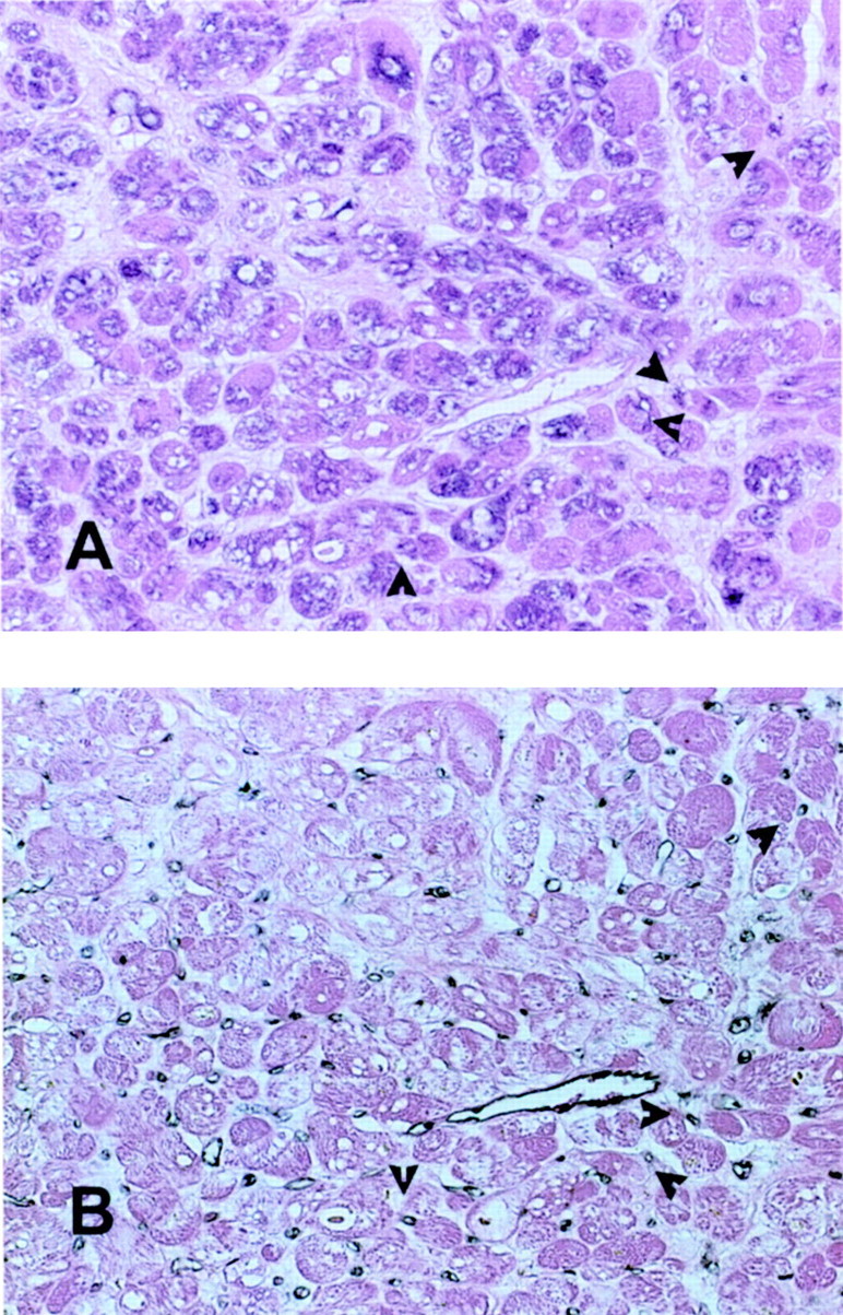
Immunohistochemical staining for MCP-1 in a myocardial segment with recovery of function after revascularization (A). Diffuse staining is noted in many cardiomyocytes and occasional microvascular endothelial cells (arrows) identified by serial section staining with a monoclonal antibody to CD31 (B). Original magnifications, ×400.