
Figure 1.
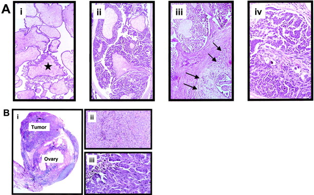
A: Morphological features of the different ovarian serous neoplasms. i: SBT with focal transition to a micropapillary area (asterisk). These focal micropapillary areas are not infrequent in SBTs. ii: Noninvasive MPSC completely replaces an ovary. The tumor is characterized by broad fibrotic papillae from which numerous long delicate micropapillary projections emanate. iii: Noninvasive MPSC (in the upper field) with an area of early invasion (invasive MPSC; right lower field with arrows). iv: Invasive MPSC. The low-grade nuclear features in the invasive carcinoma are the same as in the noninvasive counterpart. Original magnification, ×250. B: Morphological features of conventional serous carcinoma. i: Small (early) conventional serous carcinoma. The tumor measures 0.7 cm and appears to arise from the overlying ovarian surface epithelium. ii: Conventional serous carcinoma. In this area the tumor is composed of solid masses. iii: Conventional serous carcinoma. Spaces within the masses create a pseudopapillary structure as a result of cellular necrosis and processing artifact. The tumor cells contain highly atypical nuclei. Original magnification, ×400.