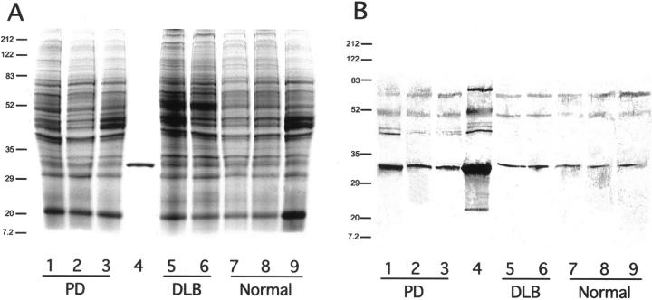
Figure 1.
A: PAGE analysis of soluble brain proteins from white and gray matter of normal, PD, and DLB brains. Lane 1, PD, FC, and GM; lane 2, PD, FC, and WM; lane 3, PD and SN; lane 4, NSGP recombinant protein; lane 5, DLB, FC, and GM; lane 6, DLB, FC, and WM; lane 7, N, FC, and GM; lane 8, N, FC, and WM; and lane 9, N and SN. Gel was stained with Coomassie blue. Molecular weights in kd are indicated at the left. Abbreviations: FC, frontal cortex; GM, gray matter; WM, white matter; SN, substantia nigra; DLB, dementia with LBs, N, normal. B: Western blot of the same samples as indicated in A. Primary antibody was protein A-purified rabbit anti-NSGP IgG, secondary antibody was a biotin-labeled donkey anti-rabbit (711-065-152, Jackson ImmunoResearch) and visualized with a Vector ABC kit using DAB.

