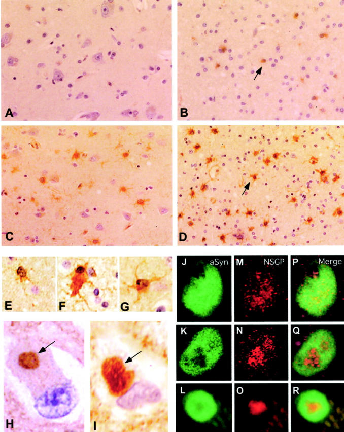
Figure 2.

A–D: Low-power images (original magnifications, ×400) of the NSGP immunocytochemistry of gray and white gray matter from normal (A and B) and DLB (C and D) brain tissue. NSGP was visualized using a biotinylated donkey anti-rabbit secondary antibody and a Vector ABC staining kit with DAB. Sections were lightly counterstained with hematoxylin. NSGP-positive glial cells were markedly increased in both gray and white matter of DLB compared to normal. The arrows in C and D indicate NSGP-positive glial cells. E–G: Comparison of astrocyte labeling (oil immersion; original magnifications, ×1000) with NSGP antibodies in normal white cortical matter (E), and white (F) and gray matter (G) of DLB cortical tissue. H and I: LB labeling with rabbit anti-NSGP antibodies (H), and labeling with sheep anti-α-synuclein antibodies (I) within the gray matter of DLB tissue (oil immersion; original magnifications, ×1000). Immunoreactivity was visualized with biotinylated secondary antibodies and a Vector ABC kit using DAB. Arrows indicate the positively stained LBs within the cytoplasm of neurons. J–R: J to L show α-synuclein labeling with fluorescein isothiocyanate in homogeneous LBs (J) and two forms of concentric LBs (K and L); M to O show NSGP labeling with Cy5 in the same three LBs; and P to R depict the merged image showing the co-localization.
