
Figure 1.
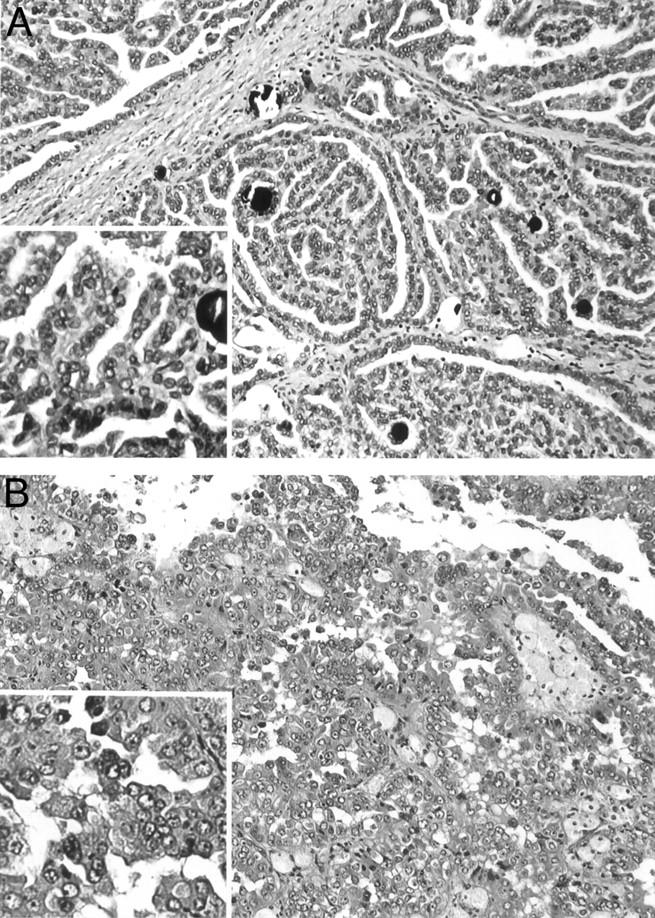
Histopathology of representative PRCC. A: Type 1 PRCC case 153 characterized by papillae covered by small tumor cells with scant pale cytoplasm and small nuclei. B: Type 2 PRCC case 173 characterized by papillae covered by large tumor cells with abundant eosinophilic cytoplasm and large nuclei with prominent nucleoli.