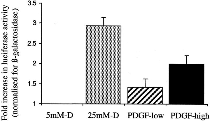
Figure 3.
Alteration in TGF-β1 promoter activity. Ninety percent confluent monolayers of HK-2 cells were transfected with the TGF-β1 promoter-luciferase plasmid construct pGL3-TGF-β1 + 11/−1362 and the control plasmid pSV-β-galactosidase before exposure to 5 mmol/L of d-glucose (control), 25 mmol/L of d-glucose (dotted bar), 25 ng/ml of PDGF-AA (hatched bar), or 100 ng/ml of PDGF-AA (solid bar). After 12 hours total cell protein was extracted and analyzed for luciferase and β-galactosidase activity. Data are expressed as luciferase activity divided by β-galactosidase activity normalized to the values obtained for control cells. Results represent mean ± SE for four individual experiments.

