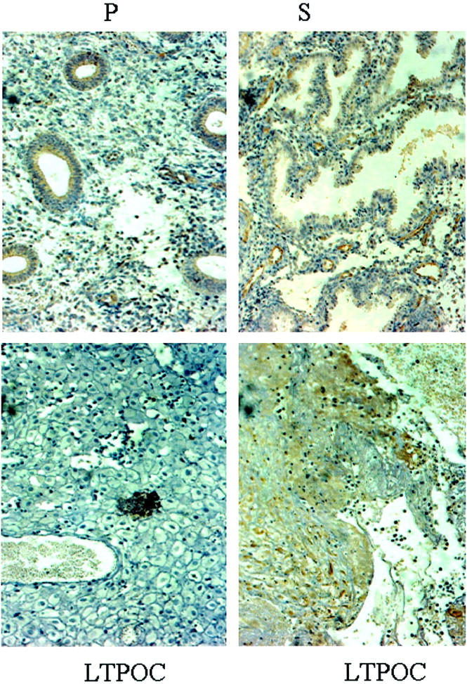
Figure 1.

Immunohistochemistry of Ang-1 in control and LTPOC-treated human endometrium. Immunohistochemical staining for Ang-1 was performed in human endometrium from proliferative phase (P), secretory phase (S), and endometrium exposed to high-dose local intrauterine LNG (LTPOC) for 3 months. Two different areas from the same pseudo-decidualized tissue show the consistent blotchy staining pattern after LTPOC treatment reflected by an area of low to no staining (bottom left) and an area of high staining (bottom right). Studies were performed in paraffin-fixed sections as described in Materials and Methods. Antigens are identified by brown peroxidase staining. No staining was observed in the absence of primary antibody (not shown).
