
Figure 6.
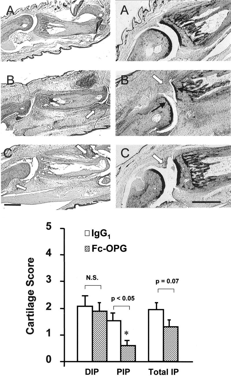
Top: Effect of Fc-OPG on cartilage. Consecutive sections show representative IP joints from normals (A), CIA treated with IgG1 (B) or CIA treated with Fc-OPG (C) for 5 days. Left: H&E stain. Right: Toluidine blue stains for cartilage. Note digital edema and synovial inflammation (arrowheads) in arthritic rats treated without or with Fc-OPG and cartilage loss (black arrows) in IgG1 controls. Bar, 500 μm. Bottom: Effect of Fc-OPG on cartilage erosion scores. The IP joints from arthritic rats treated without (n = 10) and with (n = 10) Fc-OPG were evaluated for cartilage erosions using a scoring system [as described in Materials and Methods]. Results are expressed as the mean (±SEM) scores for distal IP (DIP), proximal IP (PIP), or total IP joints of each intervention group. (*, P < 0.05).