
Figure 7.
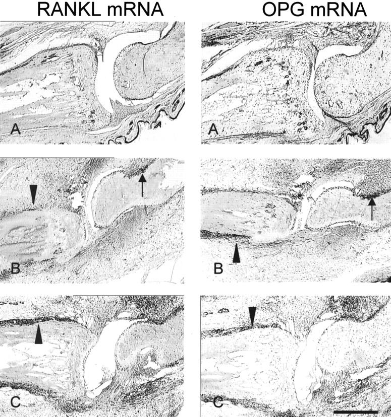
In situ localization of RANKL and OPG mRNA in CIA. Consecutive IP joint sections were processed for DIG-labeled in situ hybridization as described in Methods. A, Normal; B, CIA + IgG1; C, CIA + Fc-OPG. Positive signal for the respective mRNAs is seen as dark purple staining. Note the high RANKL and OPG mRNA expression at the sites of bone erosion (black arrows) and periosteal inflammation (arrowheads). Bar, 500 μm.