
Figure 1.
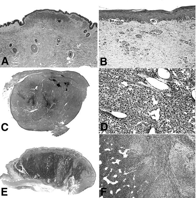
Histopathology of cases in groups II to V. A: Photomicrograph of case CN12, representative for cases of group II. In the center of the superficial dermis there is focus of increased cellularity in an otherwise bland superficial congenital nevus. B: Case CN14, representative of group III, showing a junctional melanocytic proliferation with melanocytes disposed in irregular nests and solitary units, simulating superficial spreading melanoma. This case additionally shows marked desmoplasia and irregularly configured nests of melanocytes in the dermis. Scanning magnification and close up of case D62, representative of cases in group IV (C, D), and case CN10, the only case in group V (E, F). The images show a large, sharply demarcated dermal nodule (C) with increased cellularity and marked angiogenesis (D). A large dermal nodule (E) with areas of increased cellularity and a hemangiopericytoma-like vascular pattern (F, left), and areas with spindle-shaped cells in a myxoid stroma (F, right).