Figure 2.

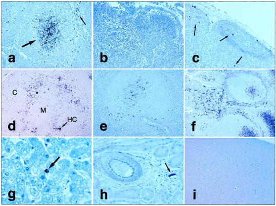
FIV chromogenic immunohistochemistry. a: FIV antigen+ cells in lymph node germinal center (large arrow) and paracortical cells (small arrow). b: No FIV antigens in sham-inoculated control cat lymph node. c: Scattered labeled cells (arrows) by RNA in situ hybridization in lymph node from FIV+ cat. FIV immunohistochemistry on tissues from FIV+ cat including: thymic lobule with clustering of antigen+ cells along junction between cortex (C) and medulla (M), with additional antigen localization in or near Hassal’s corpuscle (HC) (d, arrow); viral antigen expression in splenic periarteriolar lymphatic sheaths (e); abundant FIV antigen in ileal lamina propria leukocytes and mucosal-associated lymphoid tissue (Peyer’s patch) germinal centers (f); one antigen+ mononuclear cell (arrow) in lumen or lining hepatic sinusoid (g); apparently cell-free virus lining endothelial surface of small venule in kidney (arrow), but not in renal tubules or larger artery (at left; h); and brain with no detectable viral antigens (i). Original magnifications: ×200 (a, c, and e), ×100 (b and d), ×240 (f), ×600 (g), ×400 (h), and ×40 (i).
