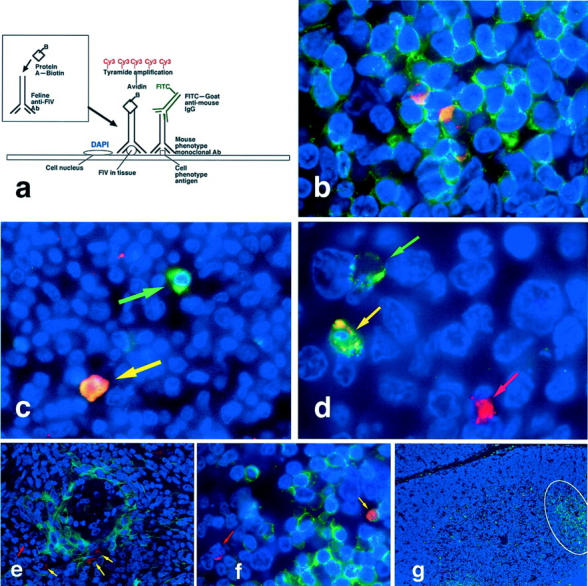
Figure 3.

Fluorescence immunohistochemistry. a: Diagram of three-color immunofluorescence assay demonstrating prelabeling of heterologous antiserum with biotin-protein A followed by tissue binding and Cy3-tyramide amplification for red labeling of FIV, fluorescein isothiocyanate-conjugated secondary antibodies for green labeling of cell phenotype antibodies, and DAPI counterstaining for blue labeling of nuclear chromatin. b: FIV antigens co-localized with CD3+ T cells in lymph node paracortex. c: An FIV+ AM-3K-labeled macrophage (yellow arrow) and a macrophage without FIV antigens (green arrow) in lymph node medulla. d: S-100+ dendritic cells with (yellow arrow) and without (green arrow) FIV antigens in lymph node follicle; also present is a FIV+ S-100− cell (red arrow). e: A few cytokeratin+ thymic epithelial cells (yellow arrows) as well as cytokeratin− FIV+ cells (red arrow) that are probably mature thymocytes in medulla. f: FIV antigens in both CD3+ (yellow arrow) and CD3− (red arrow) cortical thymocytes. g: CD45R/B220+ B cell infiltrates, occasionally forming pseudofollicles (outlined) in thymus of FIV-infected cat. Original magnifications: ×1000 (b and d), ×600 (c and f), ×400 (f), ×200 (e), and ×100 (g).
