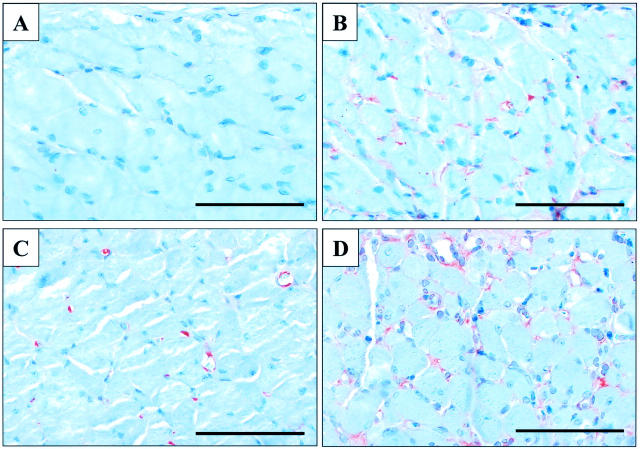
Figure 4.
Photomicrographs showing immunostaining for HIF-1α and VEGF in rat esophageal muscularis propria 3 days after sham-operation or ulcer induction. A: HIF-1α signal is absent in sections from sham-operated rats. B: HIF-1α expression in microvessels adjacent to the necrotic tissue of the ulcer bed. C: VEGF expression in microvessels of sham-operated rats. D: VEGF expression in microvessels adjacent to the necrotic tissue of the ulcer bed. Scale bars, 100 μm.

