Figure 8.

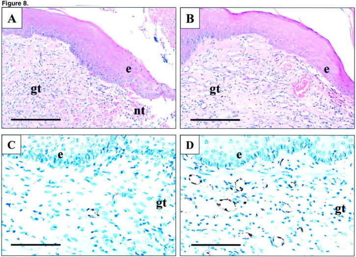
Photomicrographs of esophageal ulcer margin 7 days after injection of indicated plasmids. A and C: Control plasmid. B and D: Plasmid encoding rhVEGF165. A and B: H&E staining. C and D: Immunostaining for Factor VIII-related antigen. Factor VIII-related antigen expression (brown staining) is present in the cytoplasm of endothelial cells forming microvessels. e, epithelium; gt, granulation tissue; nt, necrotic tissue. Scale bars, 200 μm (A and B); 100 μm (C and D).
