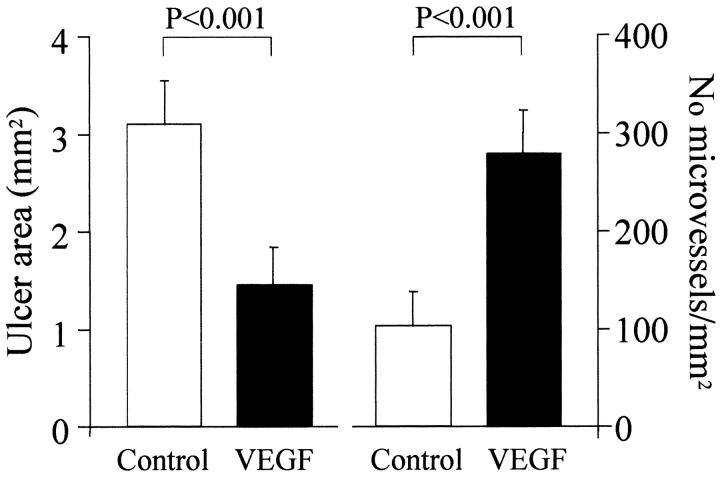
Figure 9.
Quantitative evaluation of ulcer area (left) and microvessel density in granulation tissue below the epithelium of the ulcer margin (right) in rats injected either with control plasmid (control) or plasmid encoding rhVEGF165 (VEGF) 7 days after ulcer induction/injection. Ulcer area (area of mucosal defect) was measured by a computerized video analysis system. Microvessel density was calculated as the number of microvessels per mm2 of granulation tissue section. Values are means ± SD. For each column, n = 6.

