
Figure 1.
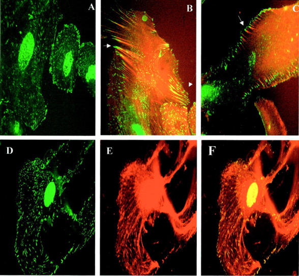
A–C: Subcellular distribution of nephrin and actin—double immunolabeling. A: Nephrin (green) expressed at cell surface and intracytoplasmically. B: Nephrin (green) double staining with AFs (red), showing nephrin cell membrane localization, and co-localization along AF extending into processes (dashed arrow), and mature expression at tips of AF (solid arrow). Cytoplasmic nephrin is seen to be distinct from AF. C: Nephrin (green) double staining with AF (red), showing mature peripheral nephrin expression at tips of AF (arrow). D–F: Nephrin co-localization with podocin. D: Nephrin cytoplasmic and cell surface distribution. E: Podocin, showing same distribution pattern as nephrin, with a more complete, filamentous appearance. F: Merged image, showing co-localization of nephrin with podocin appearing as yellow, with the rest of the podocin filaments remaining as red.