
Figure 4.
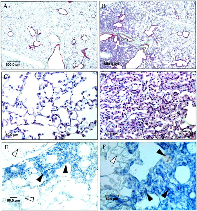
Histopathology of mice recovered for 10 days. A modified Movat stain was performed on p21-wild-type (A) and deficient lungs (B–D). C and D represent higher power views of alveolarized (C) and hyperplastic regions (D) of p21-deficient lungs. Note minimal collagen (light green) or proteoglycan (light blue) staining in A–D. p21-deficient (E, F) lungs were immunostained for α-smooth muscle actin and BrdU. α-Smooth muscle actin stained blue and BrdU stained brown. Filled arrow denotes cells expressing α-smooth muscle actin and open arrow denotes cells lacking expression.