
Figure 7.
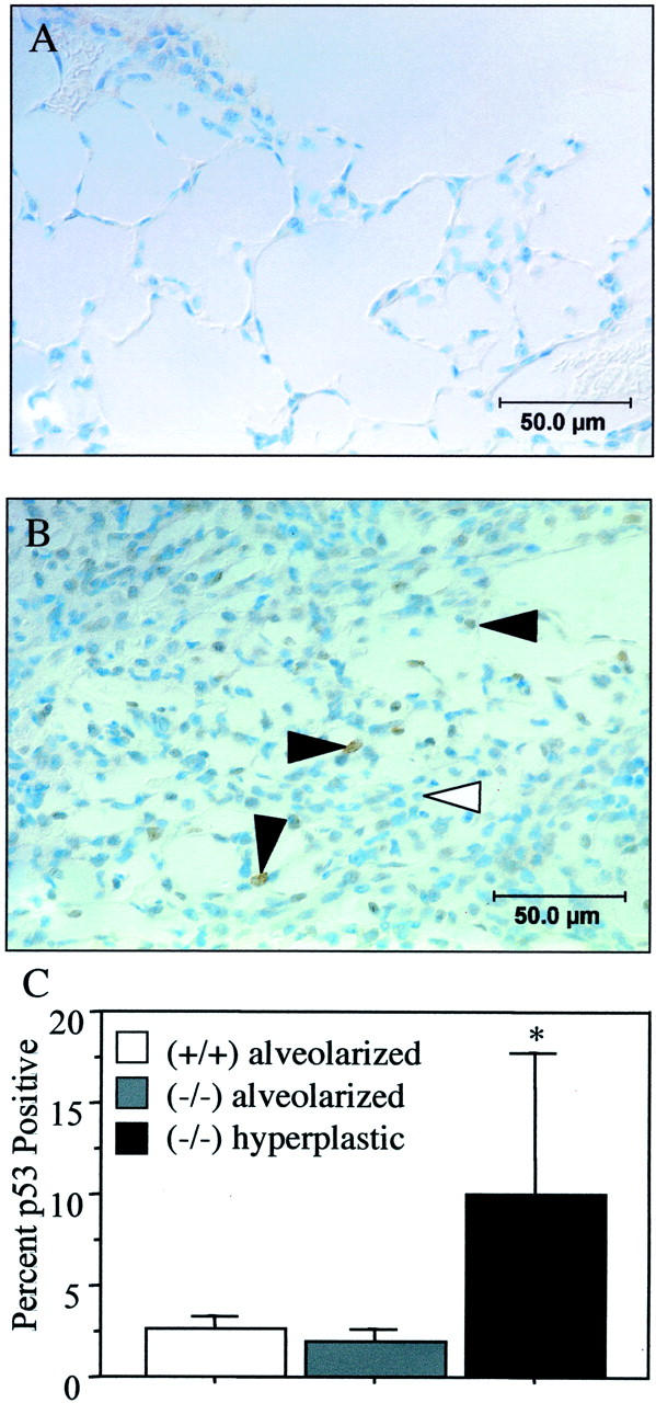
p53 expression in recovered lungs. p53 staining of p21-wild-type (A) and hyperplastic regions of p21-deficient (B) lungs recovered for 10 days. Filled arrowhead denotes cells expressing p53. C: The percentage of p53-positive cells was increased in hyperplastic regions of p21-deficient mice compared to their alveolarized regions and compared to parenchyma of p21-wild-type mice (*, P < 0.01).