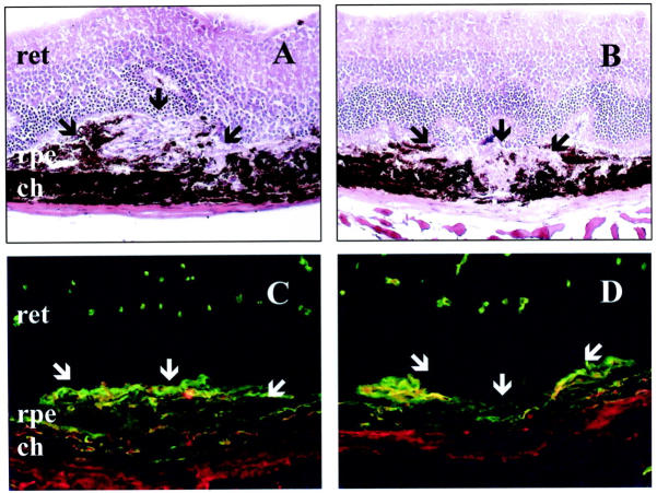
Figure 5.
Histological analysis after laser treatment. Hematoxylin-eosin staining of a representative area of choroidal neovascularization at the site of laser-induced trauma in WT mice (A), and a more restricted reaction in MMP-9−/− mice (B). Immunofluorescence labeling of new vessels in WT (C) or in MMP-9−/− mice (D) analyzed 14 days after laser photocoagulation. New vessels were detected with anti-mouse anti-collagen type IV antibody (green) and anti-mouse anti-PECAM antibody (red). The neural retina (ret), retinal pigment epithelium (rpe), and choroidal layer (ch) are indicated and the neovascular area is arrowed. White arrows localize the laser impact. Original magnification, ×200

